")
")


THE UPSHOT
Diagnostic imaging has become more common in wellness and longevity spaces, expanding beyond urgent medical use into routine check-ups, wellness centers, and spa offerings. These tools can be lifesaving, and there are important things to be aware of regarding their use.
The most empowered approach is to understand what each scan does, when it is recommended, and how it fits into a broader strategy of building resilience from within. True prevention is less about the number of scans you undergo and more about the strength of your body’s terrain, or the internal environment where health takes root.
A Shifting Landscape
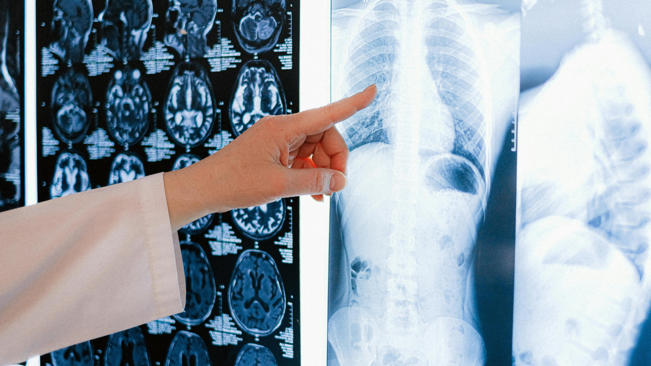
A generation ago, most people only encountered imaging technology when something was seriously wrong–a broken bone, unexplained bleeding, or a suspected tumor. Today, these tests are used in routine wellness practices. Bone-density scans are offered in wellness spas, dental offices promote advanced 3-D imaging, MRIs are prescribed for unidentified aches, CT scans for headaches, and more. These tools are saving lives. They deserve a closer look in the context of longevity, where prevention means more than just surveillance.
Scans 101
Each imaging technology uses a different method to gather information, and each carries its own benefits and limitations.
A DEXA scan, or Dual-Energy X-ray Absorptiometry, is most often used to measure bone density and increasingly, body composition. It involves very low-dose radiation, far less than most other forms of imaging (1).
An MRI, or Magnetic Resonance Imaging, uses magnets and radio waves to create detailed images of soft tissues. It does not involve radiation, making it a safer choice when repeated imaging is required.
A CT scan, or Computed Tomography, combines multiple X-rays into cross-sectional images. It is excellent for internal detail. It also delivers a higher radiation dose than a standard X-ray.
Ultrasound uses sound waves to visualize organs and blood flow. It carries no radiation risk and is commonly used for pregnancy, abdominal concerns, and vascular health.
Dental X-rays are designed to detect cavities, bone loss, and infection. While the radiation is relatively low, these can be repeated at routine cleanings (2).
A cone beam CT scan, often used in dental offices for 3-D imaging, involves higher radiation than standard dental X-rays. It can be valuable in planning implants or complex procedures and is not generally recommended for routine dental check-ups (3).
There are also unconventional or boutique offerings such as whole-body CT scans marketed as preventive, despite the high radiation dose and the benefit of the whole-body CT is more likely to be highly personalized. Whole-body MRIs are radiation-free and are significantly more expensive. Thermography is radiation free and less reliable than standard imaging.
Radiation Exposure: What’s Normal?
Radiation exposure is measured in millisieverts, or mSv. On average, we all receive about 3 mSv per year from natural background sources such as cosmic rays, soil, and sunlight. In medicine, the guiding standard is known as the ALARA principle: exposure should be kept “As Low As Reasonably Achievable.”
To put imaging in perspective, a DEXA scan delivers about 0.001 mSv, less than a single day of background exposure. An MRI and an ultrasound both involve zero radiation. A CT scan of the abdomen or pelvis, on the other hand, is closer to 10 mSv, which is the equivalent of three years’ worth of background radiation.
Dental bitewing X-rays, often taken at cleanings, deliver about 0.005 mSv, which is similar to the radiation you might absorb on a short plane flight. A full mouth series of dental X-rays comes closer to 0.15 mSv, equal to two weeks of background exposure. Cone beam CT scans can range from 0.05 to 0.5 mSv, or up to six months of background exposure. For context, a chest X-ray delivers 0.1 mSv, roughly ten days of background exposure, while a mammogram averages 0.4 mSv, equal to about seven weeks of background exposure.

How Often Are Scans Recommended?
Frequency depends on the scan, the individual’s risk profile, and professional guidelines. Recommendations are most often issued by the U.S. Preventive Services Task Force and specialty associations such as the American Dental Association.
A DEXA scan is generally advised every two years for women over 65, or earlier for those with elevated risk, like family history of osteoporosis. CT and MRI scans are usually recommended only when symptoms or physical exam findings suggest that imaging would change the treatment plan. Dental X-rays may be suggested every one to two years for those at high risk of cavities, and every two to three years or longer for those at lower risk. Routine X-rays at every cleaning are not always necessary. Mammograms are generally recommended every one to two years for women between the ages of 40 and 74, depending on individual risk factors.
Beyond the Numbers: Additional Considerations
Before undergoing imaging, you should consider benefits and costs, beyond radiation exposure. The information could be critical to ruling out positive diagnosis, and help pinpoint the most effective treatment plan.
On the other hand, don’t dismiss incidental findings. Imaging can uncover anomalies that are benign. They can lead to more testing, procedures, or even surgeries that may not improve long-term outcomes. Also, take into account the process. Preparing for scans, waiting for results, and managing uncertainty can fuel anxiety.
From an energetic perspective, repeated imaging can create a sense of disconnection from the body. The sterile medical environment, loud machines, and clinical procedures may leave a person feeling drained or unsettled. In Reiki practice, this is sometimes described as a disruption of natural flow, and it can be reflected in subtle changes in mood, sleep, or nervous-system balance.
There may also be nervous-system stress. Undergoing a scan can push the body into a fight-or-flight state. Even short exposures can leave lingering imprints on the autonomic nervous system, reducing heart-rate variability, impairing digestion, and elevating inflammation over time.
Cultivating Resilience from Within
We often talk about collecting data to further personalize treatment in order to prevent chronic conditions. In longevity, though, prevention means more than ruling out disease–it means cultivating resilience.
You may have heard the word terrain to describe the body’s internal environment: immune tone, inflammatory balance, metabolic health, microbiome diversity, sleep quality, hormone rhythms, nervous-system regulation, and social-emotional context.
When the terrain is strong, the body can self-correct small imbalances without escalating into illness. Of course, we should conduct imaging when we can glean critical data for necessary treatment. When the terrain is strong, however, the body may have all it needs to heal naturally and any image findings may simply show a body in the midst of this process.
Supporting the whole body involves:
- Eating the rainbow, a diverse, plant-forward diet rich in fiber and fermented foods to support the gut-immune axis and help manage blood sugar balance.
- Prioritizing restorative sleep and light exposure to regulate hormones.
- Building strength through resistance training to protect bone and muscle, and incorporating gentle movement into your routine.
- Practicing breathwork, meditation, or Reiki to shift nervous-system tone back toward parasympathetic balance.
- Using targeted therapies such as red-light, NAD+ support, or lymphatic drainage when they enhance recovery rather than chase biomarkers.
A Balanced Approach
Scans and imaging can save lives.The most empowered path is to know what each scan entails, how much radiation it involves, how often it is recommended, and most importantly, whether the results will meaningfully influence your care.
Ask questions, request alternatives where appropriate, and integrate practices that help restore balance afterward.
Prevention in longevity is about creating a body and mind resilient enough to thrive. By cultivating your terrain and approaching technology with discernment, you can make diagnostic imaging a powerful ally.
xo – Serena
FAQs
- Do dental wands that scan for 3-D printing expose you to radiation like X-rays?
A. No. Intraoral scanners used for digital impressions and 3-D printing rely on light, not X-rays. They capture detailed images of the teeth and gums without radiation exposure. These scans can be helpful for crowns, implants, and orthodontics but don’t replace X-rays for detecting cavities or bone loss. - What does a doctor mean when they compare a dental X-ray to “a short plane ride”?
A. Radiation during flights comes from cosmic rays at high altitude. A dental bitewing X-ray is often compared to the dose you’d get on a flight like New York to Boston. It’s a useful analogy but can be misleading, since most people take flights only occasionally while dental imaging may be repeated several times per year. - Why do MRIs or ultrasounds get recommended for minor injuries, like a bumped knee?
A. Often it’s about reassurance and liability. Imaging can quickly rule out structural problems, but in many cases rest, physical therapy, or observation would resolve the issue. Ordering a scan “just in case” may add cost and anxiety without improving recovery. - Are there tests that use radiation but are less mainstream?
A. Yes. Examples include whole-body CT scans marketed as preventive screenings, which deliver high radiation without strong evidence of benefit, and some boutique “longevity” offerings like repeated full-spine imaging or cardiac calcium scoring done too frequently. These can add up to significant radiation over time. - Besides radiation, what other costs come with unnecessary imaging?
A. Financial cost is one piece — CT scans can range from $1,000 to $5,000 out of pocket. But there’s also “mental cost.” More imaging increases the chance of incidental findings, which can lead to follow-up tests, biopsies, or procedures. Even benign results often cause weeks of anxiety. - How can imaging affect energy balance from a holistic perspective?
A. From an energetic standpoint, repeated scans may create subtle disruptions in the body’s field. Clients often describe feeling ungrounded or “out of sync” afterward. This may reflect how the parasympathetic nervous system is unsettled by the clinical process itself, or how the mind-body connection interprets repeated medical scrutiny as stress. - How can patients stay grounded if a scan is truly necessary?
A. Strategies include practicing slow breathing before and after the scan, visualizing protective light around the body, booking the appointment earlier in the day to reduce anticipatory anxiety, and following the scan with grounding activities like walking outdoors or Reiki. These small steps help rebalance both nervous-system tone and energetic flow.
CITATIONS
- To scan or not to scan? DXA in postmenopausal women | Cleveland Clinic Journal of Medicine
- radiation-safety-in-dental-practice-cda.pdf
- Mah E, Ritenour ER, Yao H. A review of dental cone-beam CT dose conversion coefficients. Dentomaxillofac Radiol. 2021 Mar 1;50(3):20200225. doi: 10.1259/dmfr.20200225. Epub 2020 Nov 11. PMID: 33112658; PMCID: PMC7923070.
by